Automated Macular Pathology Diagnosis in Retinal OCT Images using Multi-scale Spatial Pyramid and Local Binary Patterns in Texture and Shape Encoding (published in MICCAI 2010 (oral), MIA 2011, IOVS 2011)
 |
|||
| Authors | |||
|
Yu-Ying Liu1,
Mei Chen2,
Hiroshi Ishikawa3,4,
Gadi Wollstein3,
Joel S. Schuman3,4, and
James M. Rehg1
1School of Interactive Computing, Georgia Institute of Technology, Atlanta, GA 1Intel Labs Pittsburgh, Pittsburgh, PA 3UPMC Eye Center, University of Pittsburgh Medical Center, Pittsburgh, PA 4Department of Bioengineering, University of Pittsburgh, Pittsburgh, PA | |||
| Abstract | |||
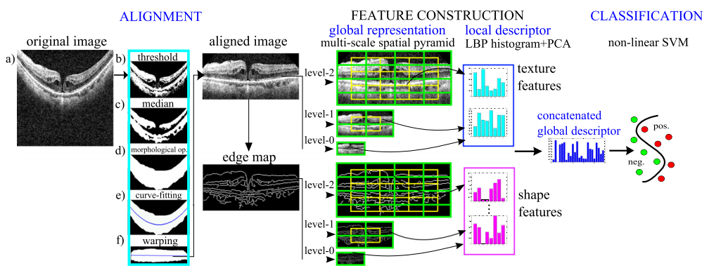
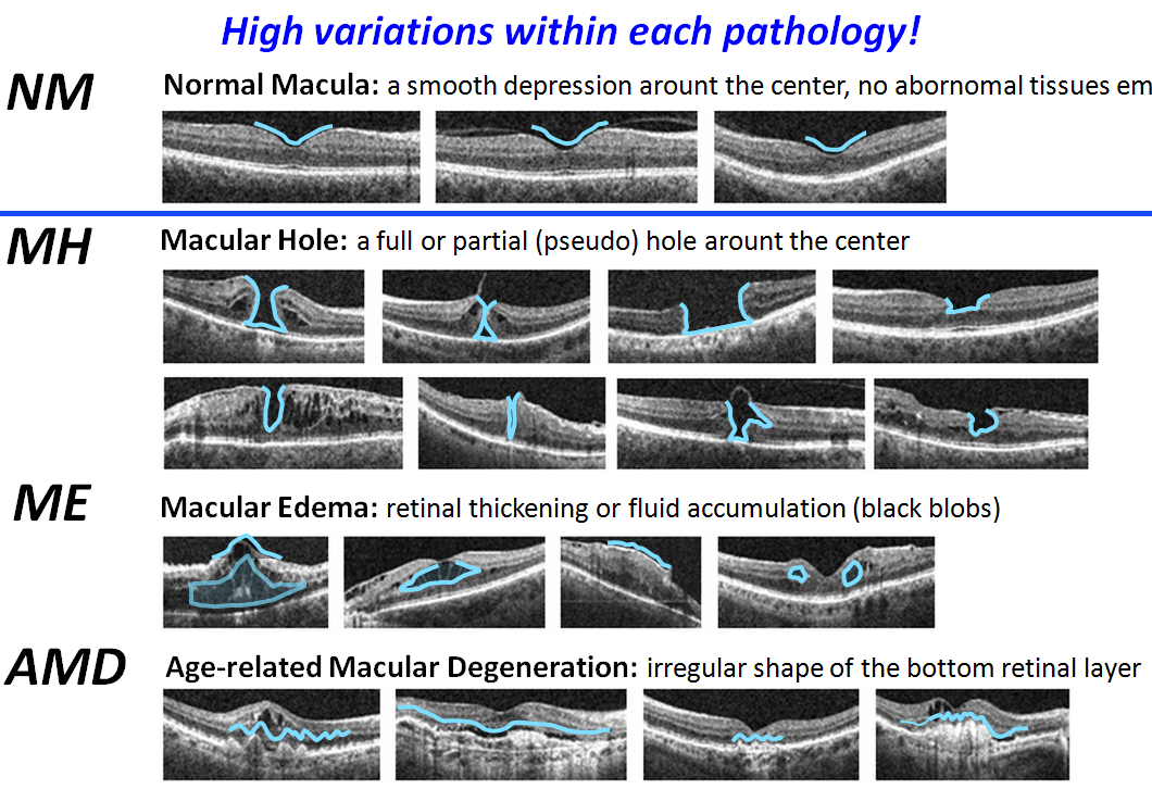
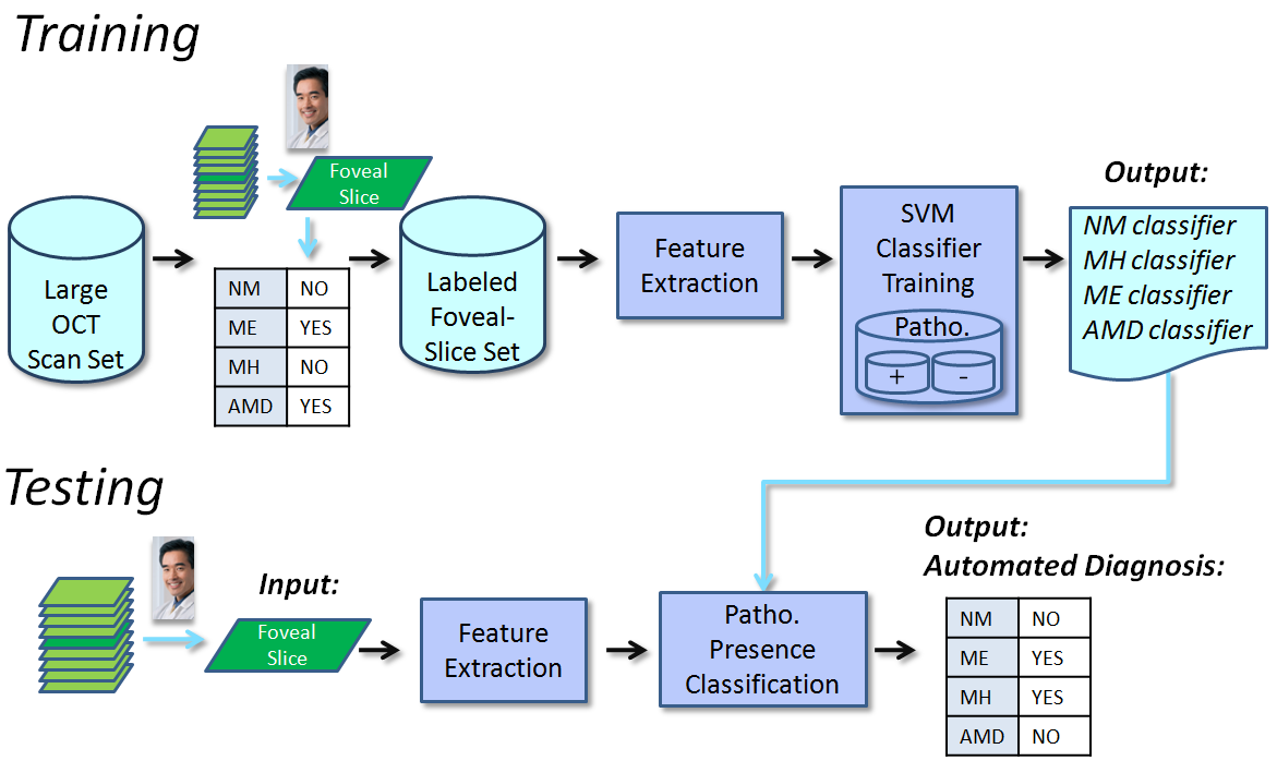
| We address a novel problem domain in the analysis of optical coherence tomography (OCT) images: the diagnosis of multiple macular pathologies in retinal OCT images. The goal is to identify the presence of normal macula and each of three types of macular pathologies, namely, macular edema, macular hole, and age-related macular degeneration, in the OCT slice centered at the fovea. We use a machine learning approach based on global image descriptors formed from a multi-scale spatial pyramid. Our local features are dimension-reduced local binary pattern histograms, which are capable of encoding texture and shape information in retinal OCT images and their edge maps, respectively. Our representation operates at mul-tiple spatial scales and granularities, leading to robust performance. We use 2-class support vector machine classifiers to identify the presence of normal macula and each of the three pathologies. To fur-ther discriminate sub-types within a pathology, we also build a classifier to differentiate full-thickness holes from pseudo-holes within the macular hole category. We conduct extensive experiments on a large dataset of 326 OCT scans from 136 subjects. The results show that the proposed method is very effective (all AUC > 0.93). | |||
| Reference | |||
|
Y.-Y. Liu , M. Chen, H. Ishikawa, G. Wollstein, J.S. Schuman, J. M. Rehg,
"Automated macular pathology diagnosis in retinal OCT Images using multi-scale spatial pyramid with local binary patterns",
Intl Conf on Medical Image Computing and Computer Assisted Intervention (MICCAI), 2010
(Oral paper, acceptance rate: 5.7% out of 786 submissions) (Finalist (Top 15) of MICCAI Young Scientist Award, 2010) [PDF] [Slides] Y.-Y. Liu , M. Chen, H. Ishikawa, G. Wollstein, J.S. Schuman, J. M. Rehg, "Automated Macular Pathology Diagnosis in Retinal OCT Images using Multi-scale Spatial Pyramid and Local Binary Patterns in Texture and Shape Encoding", Medical Image Analysis (MIA), 15 , 2011 [PDF] Y.-Y. Liu , M. Chen, H. Ishikawa, G. Wollstein, J.S. Schuman, J. M. Rehg, "Computerized Macular Pathology Diagnosis in Spectral Domain Optical Coherence Tomography Scans Based on Multiscale Texture and Shape Features", Investigative Ophthalmology and Visual Science (IOVS) , 2011 [PDF]
|
 |